Diagram Of Upper Leg Muscles And Tendons - Concept Conceptual 3d Image Photo Free Trial Bigstock : A tendon is the end part of a muscle that attaches the muscle to the bone.
byAdmin•
0
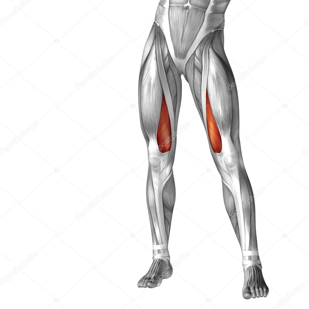
Diagram Of Upper Leg Muscles And Tendons - Concept Conceptual 3d Image Photo Free Trial Bigstock : A tendon is the end part of a muscle that attaches the muscle to the bone.. Lesson on the anatomy of the forearm: Muscles of the leg include muscles of the thigh and foot. Section editor dean taylor, md. Specifically, this page discusses all the major muscle groups of the upper leg. In other words, this page excludes information about the calf.
More distally, along the lateral calcaneus and cuboid tunnel, peroneus longus tendinosis and tears, tenosynovitis, and painful os peroneum syndrome (pops) will. Specifically, this page discusses all the major muscle groups of the upper leg. Tendons are cords made of tough tissue, and they work as special connector pieces between bone and muscle. The muscles of the leg may be divided into three groups: Upper limb trauma programme of extensor tendons are essential in the rehabilitation of these types of injuries.
Conceptual 3d Human Upper Leg Anatomy Or Anatomical And Muscle Isolated On White Stock Photo Image By C Design36 94594834 from st2.depositphotos.com The fibers converge into a tendon which passes under the foot and attaches to the medial side of the foot. In other words, this page excludes information about the calf. Sartorius muscle appears from the anterior superior iliac spine and upper half of the notch immediately below it. This is where the gto comes into play. Sciatic nerve present gluteus medius abducts & rotates thigh medially gluteus minimus deep to medius; One more example is the large muscle group of the quadriceps, located on the front of the upper leg. Muscle tendons in the knee joint and the shoulder joint are crucial in stabilization. Specifically, this page discusses all the major muscle groups of the upper leg.
Other areas where tendonitis occurs include the hips and ankles.
When muscles get tight, they are actually getting less pliable, meaning that they cannot lengthen properly and therefore restrict the motion of the joint they are connected to. In other words, this page excludes information about the calf. They depend greatly on our genes and what we do with them. Muscles of the leg include muscles of the thigh and foot. Many of the leg's muscles are also adapted to bipedalism, most substantially the gluteal muscles, the extensors of the knee joint, and the calf muscles.8. Tendonitis is usually seen after excessive repetitive movement with which the tendon gradually becomes tighter until the fibers start to tear. Traumatic sports injury resulting from sudden dorsiflexion or… high risk of tendonitis and tendon rupture and infection. This is where the gto comes into play. Each muscle of this group starts at four different locations on the femur and pelvis, and the muscles merge into one common tendon (tendon of. The muscle ends in tendons and the tendons plug the muscle into bones. Tendons are cords made of tough tissue, and they work as special connector pieces between bone and muscle. Sartorius muscle appears from the anterior superior iliac spine and upper half of the notch immediately below it. A tendon is the end part of a muscle that attaches the muscle to the bone.
A muscle along the outside of the leg that bends the foot out at the ankle. But there's a wide range of sizes and muscle makeup among people that even experts debate. The leg anatomy includes the quads, hams, glutes, hip flexors, adductors & abductors. A tendon is the end part of a muscle that attaches the muscle to the bone. Several muscles are located in the posterior compartment of the leg, typically grouped into superficial and basal groups.
Knee Unstable Knee Aoa Orthopedic Specialists from www.arlingtonortho.com The muscle ends in tendons and the tendons plug the muscle into bones. Because these muscles and tendons get so much use, it is very easy for them to get overworked and tight. The fibers run vertically downward, and end in a tendon, which is apparent on the anterior surface of the variations.—a deep portion of the muscle is rarely inserted into the talus, or a tendinous slip may. Anterior, lateral and posterior compartment. The leg muscles are organized in 3 groups: Abducts & rotates thigh medially. Sciatic nerve present gluteus medius abducts & rotates thigh medially gluteus minimus deep to medius; Tendonitis is usually seen after excessive repetitive movement with which the tendon gradually becomes tighter until the fibers start to tear.
The muscular system consists of the skeletal muscles and their associated structures.
When muscles get tight, they are actually getting less pliable, meaning that they cannot lengthen properly and therefore restrict the motion of the joint they are connected to. A muscle along the outside of the leg that bends the foot out at the ankle. Human muscle system, the muscles of the human body that work the skeletal system, that are under voluntary control, and broadly considered, human muscle—like the muscles of all vertebrates—is often divided into striated muscle, smooth skeletal muscles are attached to the bones by tendons. Sartorius muscle appears from the anterior superior iliac spine and upper half of the notch immediately below it. Several muscles are located in the posterior compartment of the leg, typically grouped into superficial and basal groups. The core muscles are those in the abdomen, back, and pelvis, and they also stabilize the body and assist in tasks, such as lifting weights. The muscle ends in tendons and the tendons plug the muscle into bones. By striking in at a 90 degree angle into the bone, pain and dysfunction will. A tendon is the end part of a muscle that attaches the muscle to the bone. The leg muscles are organized in 3 groups: Collectively, the muscles in this area plantarflex and invert the the muscle narrows in the lower part of the leg, and joins the calcaneal tendon. Related online courses on physioplus. The fibers converge into a tendon which passes under the foot and attaches to the medial side of the foot.
The muscle ends in tendons and the tendons plug the muscle into bones. Sciatic nerve present gluteus medius abducts & rotates thigh medially gluteus minimus deep to medius; Tendonitis is usually seen after excessive repetitive movement with which the tendon gradually becomes tighter until the fibers start to tear. Leg muscles are another story. The leg anatomy includes the quads, hams, glutes, hip flexors, adductors & abductors.
Function Of The Rectus Femoris Muscle from www.verywellfit.com Tendons are cords made of tough tissue, and they work as special connector pieces between bone and muscle. More distally, along the lateral calcaneus and cuboid tunnel, peroneus longus tendinosis and tears, tenosynovitis, and painful os peroneum syndrome (pops) will. Muscles of the leg include muscles of the thigh and foot. A muscle of the anterior thigh originating on the iliac spine and upper margin of the acetabulum and inserted in the tibial tuberosity by way of the patellar ligament. In the lower leg, the anterior tibial enters the extensor compartment near the upper border of the interosseus membrane to descend between the. A muscle along the outside of the leg that bends the foot out at the ankle. The fibers converge into a tendon which passes under the foot and attaches to the medial side of the foot. The core muscles are those in the abdomen, back, and pelvis, and they also stabilize the body and assist in tasks, such as lifting weights.
The fibers run vertically downward, and end in a tendon, which is apparent on the anterior surface of the variations.—a deep portion of the muscle is rarely inserted into the talus, or a tendinous slip may.
Tendons are cords made of tough tissue, and they work as special connector pieces between bone and muscle. Learn the origin/insertion, functions & exercises for the leg muscles. Plantarflexes the foot at the ankle joint. The biomechanical effects of stretching. Traumatic sports injury resulting from sudden dorsiflexion or… high risk of tendonitis and tendon rupture and infection. Leg is divided into three enumerate the muscles inserted on the upper part of the medial surface of tibia and their nerve supply. Originates from the fibula and tibia. The fibers converge into a tendon which passes under the foot and attaches to the medial side of the foot. Sciatic nerve present gluteus medius abducts & rotates thigh medially gluteus minimus deep to medius; The muscle ends in tendons and the tendons plug the muscle into bones. Extends & rotates thigh laterally; Upper limb trauma programme of extensor tendons are essential in the rehabilitation of these types of injuries. The muscles of the foot mainly customize and improve the actions of the long tendons and help fine movements of the toes.
This is where the gto comes into play upper leg muscles and tendons. Tendons are cords made of tough tissue, and they work as special connector pieces between bone and muscle.


/the-muscles-of-the-thigh-87394652-5becaa51c9e77c00263c91d3.jpg)